
Dermatological Mechanisms, Barrier Function, and Phytochemical Applications
Scope: Western dermatology, skin physiology, phytochemical mechanisms for skin support.
Table of Contents
- Skin Architecture and Barrier Function
- Wound Healing Physiology
- Inflammatory Skin Conditions
- Herbal Mechanisms: Calendula
- Herbal Mechanisms: Oats
- Clinical Applications
Skin Architecture and Barrier Function
Epidermal Structure
The epidermis is stratified squamous epithelium with four distinct layers (five in thick skin):
1. Stratum Basale (Basal Layer):
- Single layer of columnar keratinocytes
- Mitotically active (stem cells produce new keratinocytes)
- Attached to basement membrane via hemidesmosomes
- Contains melanocytes (pigment cells)
- Express keratins K5 and K14
2. Stratum Spinosum (Spinous Layer):
- Multiple layers of polyhedral keratinocytes
- Connected by desmosomes (“spines” visible under microscope)
- Begin keratin synthesis
- Switch to expressing K1 and K10
- Lamellar bodies begin forming
3. Stratum Granulosum (Granular Layer):
- 3-5 layers of flattened keratinocytes
- Contain keratohyalin granules (profilaggrin)
- Lamellar bodies mature (contain lipid precursors)
- Cornified envelope proteins synthesised (loricrin 70%, involucrin, filaggrin)
- Cells preparing for terminal differentiation
4. Stratum Corneum (Horny Layer):
- 15-30 layers of dead, flattened corneocytes
- No nucleus, no organelles
- “Brick and mortar” structure:
- Bricks: Protein-rich corneocytes (keratin + filaggrin matrix)
- Mortar: Lipid-rich extracellular matrix
- Primary barrier function
Barrier Function: The “Brick and Mortar” Model
Corneocytes (“Bricks”):
Structure:
- Flattened, dead cells
- Cornified envelope: Highly cross-linked protein shell
- Loricrin: 70% of mass
- Involucrin, cornifin, elafin, filaggrin, others
- Cross-linked by transglutaminases (gamma-glutamyl-lysine bonds)
- Extremely insoluble and resistant
Keratin filament-filaggrin matrix:
- Inside corneocyte
- Keratin intermediate filaments (K1/K10)
- Aggregated by filaggrin protein
- Provides mechanical strength
Filaggrin breakdown:
- Filaggrin eventually degraded to free amino acids
- Forms Natural Moisturising Factor (NMF)
- Exceptional water-holding capacity
- Maintains stratum corneum hydration
Lipid Matrix (“Mortar”):
Composition (ratio ~3:1:1 by weight):
- Ceramides: 40-50% (multiple subclasses)
- Cholesterol: ~25%
- Free fatty acids: 10-15%
- Small amounts of cholesterol esters
Lipid organisation:
- Arranged in lamellar sheets (multiple bilayers)
- Unique orthorhombic lattice structure
- Provides waterproofing
- Determines barrier permeability
Lipid synthesis and processing:
In stratum granulosum:
- Lamellar bodies (LBs) contain lipid precursors:
- Glucosylceramides (GlcCer)
- Sphingomyelin
- Phospholipids
- Cholesterol sulfate
- Plus enzymes: β-glucocerebrosidase, acid sphingomyelinase, phospholipases
At stratum granulosum/corneum interface:
- LBs fuse with apical plasma membrane
- Exocytose contents into extracellular space
- Enzymes process precursors:
- GlcCer → Ceramides (via β-glucocerebrosidase)
- Sphingomyelin → Ceramides (via acid sphingomyelinase)
- Phospholipids → Free fatty acids (via phospholipases)
Formation of lipid lamellae:
- Processed lipids self-assemble into lamellar sheets
- Specific ceramide subclasses critical for structure
- Particularly omega-hydroxyceramides form corneocyte lipid envelope (covalently bound to proteins)
Barrier Functions
1. Permeability Barrier:
- Prevents transepidermal water loss (TEWL)
- Without barrier: would lose ~litres of water daily
- Lipid organisation critical
- Measured by TEWL (g/mh)
Barrier disruption consequences:
- Increased TEWL
- Dehydration of deeper epidermis
- Compensatory hyperproliferation (scaling)
- Inflammatory cytokine release
- Disrupted lipid synthesis
2. Antimicrobial Barrier:
Antimicrobial peptides (AMPs):
- Produced by keratinocytes
- Secreted into stratum corneum
Major AMP families:
- Cathelicidins: LL-37 most studied
- Defensins: β-defensins (hBD-1, hBD-2, hBD-3)
- Others: Dermcidin, RNase 7, psoriasin
Mechanisms:
- Direct microbial killing (membrane disruption)
- Immune cell recruitment
- Cytokine/chemokine production
- Wound healing modulation
Dysregulation:
- Atopic dermatitis: Reduced AMP production → increased infections
- Psoriasis: Excessive AMP production → inflammation
3. Immunological Barrier:
Skin immune cells:
- Keratinocytes: Produce cytokines, AMPs; express pattern recognition receptors
- Langerhans cells: Dendritic cells in epidermis; capture antigens
- T cells: Resident memory T cells patrol epidermis
- Mast cells: Dermis; allergic responses
Immune surveillance:
- Continuous monitoring for pathogens
- Rapid local response
- Antigen presentation to adaptive immunity
Wound Healing Physiology
Phases of Cutaneous Wound Healing
Phase 1: Hemostasis (Immediate, minutes-hours)
Vascular response:
- Vasoconstriction (limits blood loss)
- Platelet activation and aggregation
- Clot formation (fibrin mesh)
Growth factor release:
- Platelets release:
- Platelet-derived growth factor (PDGF)
- Transforming growth factor-β (TGF-β)
- Vascular endothelial growth factor (VEGF)
- Initiates healing cascade
Phase 2: Inflammation (Hours to days 3-5)
Purpose: Clean wound, prevent infection, initiate repair
Cellular infiltration:
Early (hours):
- Neutrophils arrive (via chemotaxis)
- Phagocytose bacteria, debris
- Release reactive oxygen species (ROS)
- Secrete proteases
Later (days 2-3):
- Monocytes/macrophages arrive
- More efficient phagocytosis
- Secrete growth factors (PDGF, TGF-β, VEGF, FGF)
- Transition to proliferation phase
Cytokine profile:
- Pro-inflammatory: IL-1, IL-6, TNF-α
- Recruit immune cells
- Activate fibroblasts and keratinocytes
Prolonged inflammation = chronic wound
Phase 3: Proliferation (Days 4-21)
Three concurrent processes:
A. Granulation Tissue Formation:
Components:
- New extracellular matrix (ECM)
- New blood vessels (angiogenesis)
- Fibroblasts, macrophages
Fibroblast activity:
- Migrate into wound bed
- Proliferate
- Synthesise ECM:
- Collagen (type III initially, then type I)
- Fibronectin
- Hyaluronic acid
- Proteoglycans
- Differentiate to myofibroblasts (wound contraction)
Angiogenesis:
- Stimulated by VEGF, FGF
- Endothelial cells sprout from existing vessels
- Form new capillary networks
- Provides oxygen and nutrients
Appearance: Pink, granular tissue filling wound bed
B. Re-epithelialisation (Epithelialisation):
Keratinocyte response to injury:
Within hours:
- Basal keratinocytes at wound edge activated
- Change from proliferative to migratory phenotype
- Dissolve desmosomes, hemidesmosomes
- Extend lamellipodia (cellular projections)
Keratinocyte migration:
- Migrate across wound bed
- Follow fibrin/fibronectin matrix
- Keratins switch: K5/K14 → K6/K16/K17 (wound healing keratins)
- Produce matrix metalloproteinases (MMPs) to remodel ECM
Proliferation:
- Behind migrating “tongues,” keratinocytes proliferate
- Ensure adequate cell supply
- Restore epithelial thickness
Basement membrane reformation:
- Keratinocytes secrete new basement membrane components
- Re-establish hemidesmosomes
- Return to normal phenotype
Completion: When migrating fronts meet at wound centre
C. Wound Contraction:
Mechanism:
- Myofibroblasts (specialised fibroblasts with contractile apparatus)
- Express α-smooth muscle actin
- Contract, pulling wound edges together
- Reduces wound surface area
Importance: Particularly important in large wounds
Phase 4: Remodeling (Weeks to months-years)
Collagen reorganisation:
- Type III collagen → Type I collagen
- Random orientation → organised parallel bundles
- Increased tensile strength
Scar maturation:
- Cellularity decreases
- Vascularity decreases
- ECM matures
- Scar flattens, softens
Tensile strength:
- Never reaches 100% of unwounded skin
- Maximum ~80% after 1 year
- Collagen cross-linking continues improving strength
Matrix metalloproteinases (MMPs) vs. Tissue inhibitors of metalloproteinases (TIMPs):
- Balance determines ECM remodeling
- Chronic wounds: Excess MMPs, degradation exceeds synthesis
- Hypertrophic scars: Excess synthesis
Factors Affecting Wound Healing
Local factors:
- Moisture (moist wounds heal faster)
- Oxygenation
- Infection/bioburden
- Foreign bodies
- Mechanical stress
Systemic factors:
- Age (elderly heal slower)
- Nutrition (protein, vitamins C, A, zinc)
- Chronic diseases (diabetes, vascular disease)
- Medications (corticosteroids, immunosuppressants)
- Smoking (vasoconstriction, reduced oxygen)
Inflammatory Skin Conditions
Atopic Dermatitis (Eczema)
Pathophysiology:
Barrier dysfunction:
- Filaggrin deficiency: Common genetic mutation (loss-of-function FLG gene)
- Reduced NMF → Dry skin
- Impaired lipid organisation
- Increased TEWL
- Enhanced penetration of allergens, irritants
Immune dysregulation:
- Th2-predominant response
- Elevated IgE
- Cytokine profile:
- IL-4, IL-13 (drive Th2, inhibit filaggrin/lipid synthesis)
- IL-31 (pruritogenic — causes itching)
- TSLP (thymic stromal lymphopoietin)
Inflammation:
- Infiltration of T cells, eosinophils
- Mast cell degranulation
- Histamine release → Itching
- Chronic inflammation → Lichenification (skin thickening)
Itch-scratch cycle:
- Itching → Scratching → Barrier disruption → More inflammation → More itching
Microbial colonisation:
- Staphylococcus aureus overgrowth common
- Produces toxins → Superantigen effect
- Exacerbates inflammation
Herbal relevance:
Barrier support:
- Emollients (oil-based preparations) restore lipids
- Calendula, oats provide protective film
Anti-inflammatory:
- Calendula triterpenoids, oat avenanthramides reduce inflammation
Anti-pruritic:
- Oats inhibit histamine release (avenanthramides)
- Reduces itch-scratch cycle
Psoriasis
Pathophysiology:
Hyperproliferation:
- Keratinocyte turnover accelerated (~3-4 days vs. normal ~28 days)
- Incomplete differentiation
- Parakeratosis (retained nuclei in stratum corneum)
Immune activation:
- Th1 and Th17-predominant
- Cytokines:
- TNF-α, IFN-γ (Th1)
- IL-17, IL-22 (Th17)
- Drive keratinocyte proliferation
Inflammation:
- Dermal infiltration: T cells, dendritic cells, neutrophils
- Epidermal infiltration: Neutrophils form microabscesses (Munro’s)
Vascular changes:
- Dilated, tortuous capillaries in dermal papillae
- Auspitz sign (bleeding when scales removed)
Genetics:
- Strong genetic component
- HLA-Cw6 association
Herbal relevance:
Limited efficacy as monotherapy for moderate-severe psoriasis
Supportive role:
- Calendula: Anti-inflammatory, may reduce plaques
- Aloe: Soothing, mild anti-inflammatory
- Usually requires medical management
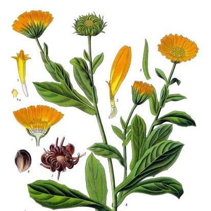
Calendula: Phytochemistry and Mechanisms
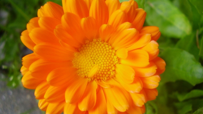
Detailed Phytochemical Profile
Triterpenoid Saponins:
Major compounds:
- Oleanolic acid glycosides
- Calendulosides A, B, C, D
- Faradiol esters
- Taraxasterol
Properties:
- Amphiphilic (both water- and fat-soluble components)
- Soap-like (saponin = “soap”)
- Hemolytic (lyse red blood cells) at high concentrations — why internal use limited
- Primary wound-healing actives
Triterpenoid Alcohols:
Major compounds:
- Faradiol: Most studied
- Taraxasterol
- α-Amyrin, β-Amyrin
- Lupeol
Properties:
- Anti-inflammatory
- Anti-oedema (reduce swelling)
Flavonoids:
Major flavonoids:
- Quercetin and derivatives
- Isorhamnetin glycosides
- Rutin (quercetin-3-O-rutinoside)
- Narcissin
Properties:
- Antioxidant (scavenge free radicals)
- Anti-inflammatory
- Antimicrobial
Carotenoids:
Major carotenoids:
- Lutein
- Zeaxanthin
- β-Carotene
- Lycopene
Properties:
- Antioxidants
- Orange/yellow pigments
- Protect healing tissues from oxidative damage
Polysaccharides:
Structure: Complex carbohydrates
Properties:
- Immunomodulatory
- Promote tissue regeneration
Volatile Oils:
Minor component but contributes antimicrobial activity
Wound Healing Mechanisms
Mechanism 1: Enhanced Granulation Tissue Formation
Triterpenoid saponin effects:
Stimulate fibroblast proliferation:
- Increase fibroblast numbers in wound bed
- Earlier formation of granulation tissue
Enhance collagen synthesis:
- Increase collagen production by fibroblasts
- Particularly type I and type III collagen
- Faster development of tensile strength
Mechanism: Likely involves growth factor signaling (TGF-β, PDGF pathways)
Mechanism 2: Promotion of Epithelialisation
Accelerates re-epithelialisation:
Keratinocyte proliferation:
- Increases keratinocyte numbers
- Faster restoration of epithelial coverage
Keratinocyte migration:
- Enhances migratory capacity
- Speeds closure of wound
Clinical evidence:
- Multiple studies show faster wound closure times with calendula
- Reduced healing time in cesarean wounds, venous leg ulcers, radiation dermatitis
Mechanism 3: Anti-Inflammatory Activity
Primary mechanism: Lipoxygenase (LOX) inhibition
Faradiol is key compound:
- Inhibits 5-LOX and 12-LOX enzymes
- These enzymes produce leukotrienes (inflammatory mediators)
- Reduced leukotriene production → Reduced inflammation
Comparative efficacy:
- Studies show faradiol more potent than indomethacin (NSAID) in some inflammatory models
- Particularly effective for edema (swelling)
Additional anti-inflammatory mechanisms:
- Flavonoids inhibit cyclooxygenase (COX) enzymes
- Reduce prostaglandin production
- Antioxidants reduce oxidative stress-induced inflammation
Result: Reduced redness, swelling, pain in inflamed/wounded skin
Mechanism 4: Antimicrobial Activity
Broad-spectrum activity:
Antibacterial:
- Effective against Gram-positive: Staphylococcus aureus, Streptococcus spp.
- Effective against Gram-negative: E. coli, Pseudomonas aeruginosa
- Includes some antibiotic-resistant strains
Antifungal:
- Candida albicans
- Dermatophytes
Mechanisms:
- Flavonoids and volatile oils disrupt microbial cell membranes
- Saponins lyse membranes (detergent-like action)
- Prevents wound infection
Mechanism 5: Antioxidant Effects
Carotenoids and flavonoids:
- Scavenge reactive oxygen species (ROS)
- Protect healing tissues from oxidative damage
- Oxidative stress impairs healing — antioxidants ameliorate
Lipid peroxidation inhibition:
- Prevents oxidative damage to lipid membranes
- Maintains cellular integrity
Bioavailability and Formulation
Topical application:
Absorption:
- Lipophilic compounds (triterpenoids) penetrate stratum corneum well
- Hydrophilic compounds (polysaccharides) less penetration
- Oil-based preparations optimal for skin delivery
Formulation considerations:
- Infused oil: Extracts lipophilic compounds (triterpenoids, carotenoids)
- Alcohol tincture: Extracts both lipophilic and hydrophilic (flavonoids, saponins)
- Aqueous extract: Polysaccharides, flavonoid glycosides
Optimal for wound healing:
- Oil-based salves/creams (lipophilic compounds penetrate skin)
- Provide occlusion (maintains moisture)
Oat Mechanisms: Anti-Pruritic Science
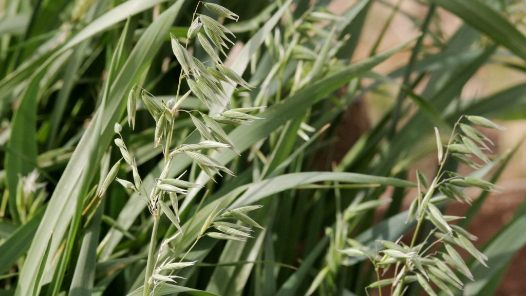
Avenanthramides: Unique Oat Compounds
Structure:
- Phenolic alkaloids
- Unique to oats (Avena species)
- Three major forms: Avenanthramide A, B, C
Anti-inflammatory mechanisms:
1. Histamine Release Inhibition:
- Directly inhibit mast cell degranulation
- Reduced histamine release
- Histamine is primary pruritogenic (itch-causing) mediator
- Result: Less itching
2. Pro-inflammatory Cytokine Suppression:
- Reduce production of:
- TNF-α (tumor necrosis factor-alpha)
- IL-6, IL-8 (interleukins)
- Mechanism: Inhibit NF-κB pathway (master inflammatory regulator)
- Result: Reduced inflammation
3. Nitric Oxide Synthase Inhibition:
- Reduce NO production in inflamed tissues
- NO contributes to vasodilation and inflammation
- Result: Reduced redness, inflammation
Beta-Glucans: Protective Polysaccharides
Structure:
- Long-chain polysaccharides (glucose polymers)
- β-1,3 and β-1,4 linkages
Mechanisms:
1. Film Formation:
- Form protective hydrocolloid film on skin surface
- Physical barrier against irritants
- Prevents allergen penetration
2. Moisture Retention:
- Hygroscopic (attract and hold water)
- Hydrate stratum corneum
- Improve barrier function
3. Immunomodulation:
- Activate pattern recognition receptors (Dectin-1)
- Enhance wound healing
- Support barrier repair
Saponins: Gentle Cleansing
Properties:
- Natural surfactants
- Gentle cleansing without stripping lipids
- Less irritating than synthetic detergents
Clinical Evidence
Atopic dermatitis studies:
- Colloidal oatmeal baths reduce itching significantly
- Improve skin barrier (reduced TEWL)
- Reduce need for topical steroids in some studies
Mechanism of bath action:
- Fine particles adhere to skin
- Form protective film
- Deliver anti-inflammatory compounds directly
Clinical Applications and Protocols
Atopic Dermatitis Management
Phase 1: Acute Flare (Days 1-7)
Topical:
- Oat baths: Daily
- Calendula cream: 3-4x daily on damp skin (occludes moisture)
- Chamomile compresses: For severe inflammation, 2-3x daily
- Avoid irritants: Harsh soaps, hot water, wool
Internal:
- Nettle infusion: 3 cups daily (anti-inflammatory, antihistamine)
- Calendula tea: 2 cups daily
- Chamomile tea: 2 cups daily (calming, anti-inflammatory)
Phase 2: Maintenance (Ongoing)
Topical:
- Oat bath: 2-3x weekly
- Emollient/calendula cream: 2x daily minimum (more if dry)
- Gentle cleansing
Internal:
- Nettle infusion: 2 cups daily
- Identify and eliminate triggers (food allergies common)
- Evening primrose oil: 1000mg daily (gamma-linolenic acid supports barrier)
Lifestyle:
- Cotton clothing
- Control environment (humidifier in winter, avoid overheating)
- Stress management
- Adequate sleep
Timeline: Improvement usually 2-4 weeks; full control may take 2-3 months
Wound Healing Protocol
Clean wounds only (no active infection requiring antibiotics)
Immediate (Day 1):
- Clean wound thoroughly (water, gentle soap if needed)
- Apply: Fresh plantain poultice OR honey OR calendula cream
- Cover with sterile dressing
Days 2-14 (until epithelialisation complete):
- Clean gently daily
- Apply calendula salve 2-4x daily
- Keep moist (moist wounds heal faster)
- Change dressing daily
Remodeling phase (Weeks 2-months):
- Continue calendula application
- May reduce to 1-2x daily
- Massage gently (once epithelialised) to reduce scarring
Infection monitoring:
- Increasing redness, warmth
- Purulent drainage
- Red streaks
- Fever
- → See doctor immediately
Safety Considerations
Calendula:
- Very safe topically
- Allergic reactions possible (Asteraceae allergy)
- Always patch test
- Internal use limited (saponins hemolytic at high doses)
Comfrey:
- Contains pyrrolizidine alkaloids (hepatotoxic)
- External use only
- Not on deep or infected wounds
- Not on broken skin for extended periods
Plantain:
- Extremely safe
- Can use liberally
Oats:
- Very safe
- Use plain oats (no added sugars/flavours)
References
Bone, K., & Mills, S. (2013). Principles and practice of phytotherapy: Modern herbal medicine (2nd ed.). Churchill Livingstone.
Guo, S., & DiPietro, L.A. (2010). Factors affecting wound healing. Journal of Dental Research, 89(3), 219-229.
Pazyar, N., Yaghoobi, R., Rafiee, E., Mehrabian, A., & Feily, A. (2013). Skin wound healing and phytomedicine: A review. Skin Pharmacology and Physiology, 26(6), 303-310.
Proksch, E., Brandner, J.M., & Jensen, J.M. (2008). The skin: An indispensable barrier. Experimental Dermatology, 17(12), 1063-1072.
Sur, R., Nigam, A., Grote, D., Liebel, F., & Southall, M.D. (2008). Avenanthramides, polyphenols from oats, exhibit anti-inflammatory and anti-itch activity. Archives of Dermatological Research, 300(10), 569-574.
Disclaimer: Does not represent rongoā Māori methods. For rongoā knowledge, consult Te Paepae Motuhake.
Medical Disclaimer: This guide is for educational purposes only and is not medical advice. Serious wounds, persistent skin conditions, changing moles, or signs of infection require professional medical evaluation. Herbal skin therapies support healing but do not replace dermatological care. Consult qualified healthcare practitioners, especially if pregnant, nursing, taking medications, or having medical conditions.
Note on Pricing: All prices mentioned in this guide are approximate and based on New Zealand suppliers as of December 2025. Prices vary by supplier, season, and market conditions. We recommend checking current prices with your local suppliers.
